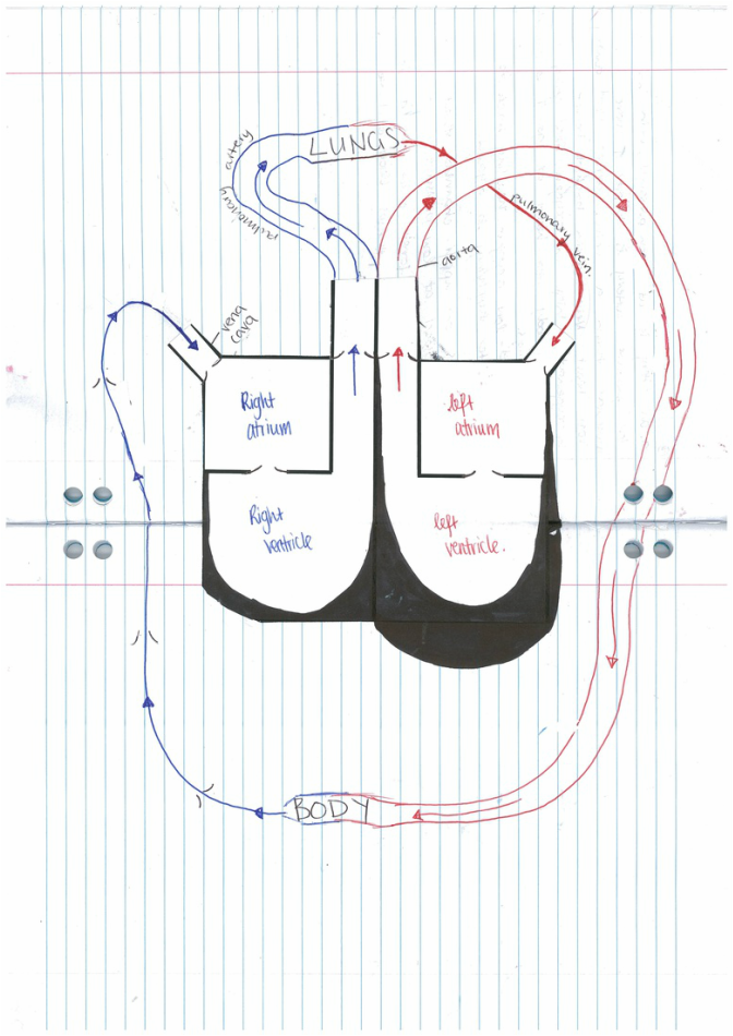
box diagram of heart
booklet questions
- Find the blood vessels on the surface of the heart muscle. These are the coronary arteries. They carry nutrients and oxygen to the heart muscle.
It is wiggly and separate out into an intricate web that spreads over the outside of the heart.
What do you think would happen if this artery was blocked by a clot?
The heart would not be able to function properly because it would not have enough oxygen.
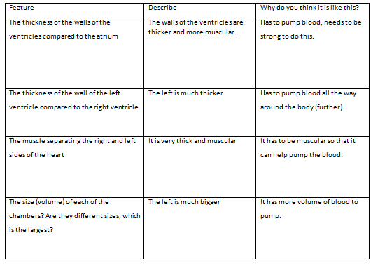
5. Have a feel of the thickness of the heart muscle at the top and bottom of the heart. Describe the following features;
a. The thickness of the muscles at the top of the heart.
They are thinner than the bottom of the heart
b. The thickness of the muscles at the bottom of the heart.
They are very thick, in particular the left ventricle.
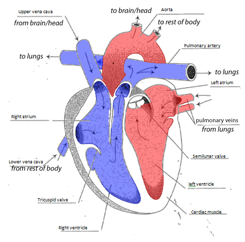
6. Find the pulmonary artery that leaves the right ventricle.
Find the pulmonary veins that enter the left atrium.
Circle the correct answer;
Oxygenated / Deoxygenated blood leaves the right ventricle / right atrium in an artery / vein and travels to the lungs / rest of the body. Here, the blood collects oxygen / drops off oxygen, so it is now oxygenated/ deoxygenated. The blood travels back to the heart via an artery / vein.
7. Find the aorta that carries the blood away from the left ventricle of the heart.
a. Describe the thickness of this vessel. Why do you think it needs to be so thick?
It is very thick and a little elastic so that it can cope with the high pressure that the blood is flowing at.
b. Where is it taking blood to?
The organs and systems of the body to supply them with oxygen.
8. Find the vena cava. This is the vein that returns blood from the body.
a. Compare the thickness of the vena cava to the aorta. Why do you think it is different?
The vena cava is thinner than the aorta because it is a vein and the blood is travelling at a low pressure it doesn’t need to be as strong to cope.
b. What part of the heart does the vena cava go back in to?
The upper right of the right atrium.
Firstly cut open the LEFT VENTRICLE following the lines on the diagram above.
2. Observe any valves you see. What do you think their job would be?
They are located between the chambers of the heart and along the vena cava. They make sure blood flows in the correct direction and there is no backflow.
Now you can cut open the RIGHT VENTRICLE following the lines on the diagram above. Observe this side of the heart.
Cut into the atria of both the left and right chambers so you can see the muscle thickness of all four chambers.
2. Fill out the table below;
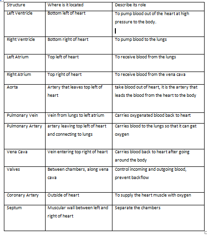
3. In the table below,
indicate the location and role of the following structures;
6.
walking the heart
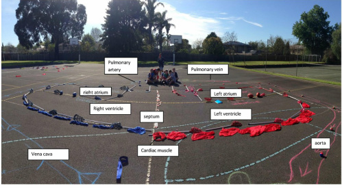
WALKING THE HEART
In the walking the heart task we used lacrosse sticks as the walls of the heart, colour coding red and blue for oxygenated and deoxygenated blood. We used pink sashes as the septum, hockey sticks and footballs as valves and blue and red bibs as cardiac muscle. We stretched the left out to show it is more muscular. For the veins we used blue chalk and drew it thinly to show that they are thinner than arteries which we drew in red chalk. We used arrows to show the direction of blood flow.
In the walking the heart task we used lacrosse sticks as the walls of the heart, colour coding red and blue for oxygenated and deoxygenated blood. We used pink sashes as the septum, hockey sticks and footballs as valves and blue and red bibs as cardiac muscle. We stretched the left out to show it is more muscular. For the veins we used blue chalk and drew it thinly to show that they are thinner than arteries which we drew in red chalk. We used arrows to show the direction of blood flow.
